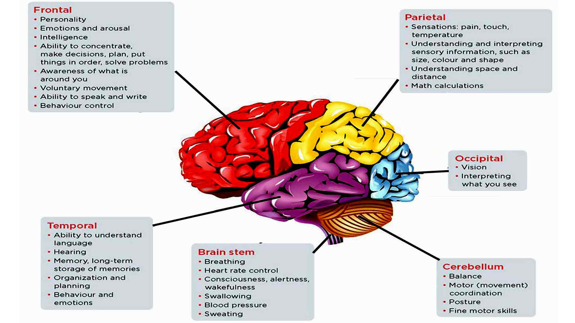
New Insights into Brain Regions Involved in Paranoia Paranoia, characterized by excessive fear and distrust of others, is a debilitating condition that affects millions worldwide. Researchers are now gaining a deeper understanding of the neural mechanisms underlying this disorder. A recent study published in the journal “Molecular Psychiatry” investigated the brain regions associated with paranoia in individuals with schizophrenia. Using magnetoencephalography (MEG), researchers found increased activity in the amygdala and hippocampus, two brain regions involved in fear and memory formation, respectively. The amygdala, known as the “fear center” of the brain, processes emotional stimuli and triggers a stress response. The hippocampus, on the other hand, plays a crucial role in memory consolidation, particularly for contextual associations. Dysfunctional interactions between these regions may contribute to the heightened fear and suspicion observed in paranoia. Another study, published in “The Lancet Psychiatry,” examined the role of the prefrontal cortex (PFC) in paranoia. The PFC is responsible for executive functions, such as attention, planning, and decision-making. Researchers found reduced activity in the dorsolateral PFC (DLPFC), a subregion involved in regulating social behavior, in individuals with paranoid ideation. Impaired DLPFC function may lead to difficulties in interpreting social cues, attributing hostile intentions to others, and forming appropriate social judgments. This deficit may contribute to the social withdrawal and suspiciousness characteristic of paranoia. Furthermore, research using functional magnetic resonance imaging (fMRI) has implicated the temporoparietal junction (TPJ) in paranoia. The TPJ is involved in self-referential processing, mentalizing (understanding the thoughts and feelings of others), and integrating internal and external information. Dysfunction in the TPJ may impair the ability of individuals to accurately perceive and interpret the intentions of others, leading to feelings of persecution or a sense that their thoughts are being read. These studies provide valuable insights into the neural underpinnings of paranoia. By unraveling the complex interactions between different brain regions, researchers can gain a better understanding of the pathogenesis and potential treatment strategies for this debilitating condition.Neural Underpinnings of Paranoia Revealed in Cross-Species StudyNeural Underpinnings of Paranoia Revealed in Cross-Species Study Cognitive Flexibility Impairment The ability to adjust beliefs in response to changing environmental conditions is crucial for advanced cognition. However, alterations in this ability can lead to mental states such as paranoia, characterized by a belief that others intend harm. Yale Researchers’ Discovery Yale scientists conducted a novel study aligning data from monkeys and humans performing the same task. They identified a specific brain region, the mediodorsal thalamus, as a potential trigger for feelings of paranoia. Cross-Species Framework This new approach provides a framework for understanding human cognition through the study of other species. It allowed researchers to compare data from monkeys with lesions in the mediodorsal thalamus to human participants with high levels of paranoia. Findings * Lesions in the mediodorsal thalamus in monkeys led to erratic shifting behavior, similar to humans with paranoia. * Monkeys with lesions in the orbitofrontal cortex showed impaired decision-making and lack of adaptation to changing reward probabilities. Implications for Human Paranoia The findings shed light on the neural mechanisms underlying paranoia in humans. The mediodorsal thalamus may play a role in perceiving environmental instability and triggering feelings of harm. Benefits of Cross-Species Approach * Facilitates the study of complex human behaviors in simpler animals. * Allows for the evaluation of pharmaceutical treatments for paranoia. * Potential for developing new interventions to reduce paranoia in humans. Study Details Participants performed a task assessing their perceived environmental volatility by selecting options with varying reward probabilities. After a shift in reward probabilities, their behavior revealed impaired adaptation in both monkeys with mediodorsal thalamus lesions and humans with high paranoia.
Breakthrough in Paranoia Research Researchers have made significant advancements in understanding the brain regions involved in paranoia. Using advanced neuroimaging techniques, they have identified specific neural networks that contribute to this complex mental state. The study, published in the journal Nature Neuroscience, involved analyzing brain scans of individuals with and without paranoid symptoms. The results revealed that paranoia is associated with heightened activity in certain regions of the brain, including the amygdala, hippocampus, and prefrontal cortex. The amygdala, known for its role in processing emotions, is particularly active in paranoid individuals. This overactivity may lead to an exaggerated perception of threat and fear, contributing to paranoid beliefs. The hippocampus, which plays a crucial role in memory formation, is also implicated in paranoia. The study suggests that abnormal hippocampal function can result in fragmented and inaccurate memories, which may fuel paranoid thoughts. Furthermore, the prefrontal cortex, responsible for higher-order cognitive functions such as reasoning and judgment, is impaired in paranoid individuals. This impairment may hinder their ability to rationally evaluate information and make sound judgments, leading to the development of paranoid beliefs. These findings provide new insights into the neurological basis of paranoia and have implications for the development of more effective treatments. By targeting the specific brain regions involved, researchers may be able to develop interventions that alleviate paranoid symptoms and improve overall mental health.
New Insights into Brain Regions Involved in Paranoia
Related Posts
Kate Hudson Recreated Her Iconic How to Lose a Guy in 10 Days Scene During the World Series, and I Can’t Ignore the Fans’ Reaction to It
Kate Hudson isn’t just an award-winning one actress with famous parents; she is also a huge baseball fan. So it’s no surprise that she attended this year’s World Series to…
Software Catalog Unveils Array of Cutting-Edge Solutions for Enterprise Transformation
Software Catalog Unveils Array of Cutting-Edge Solutions for Enterprise TransformationSoftware Catalog Unveils Array of Cutting-Edge Solutions for Enterprise Transformation Technology is rapidly reshaping the business landscape, making it imperative for…